Cierre angular agudo como una presentación atípica de cisticercosis ocular, un reporte de caso
Barra lateral del artículo
Contenido principal del artículo
Resumen
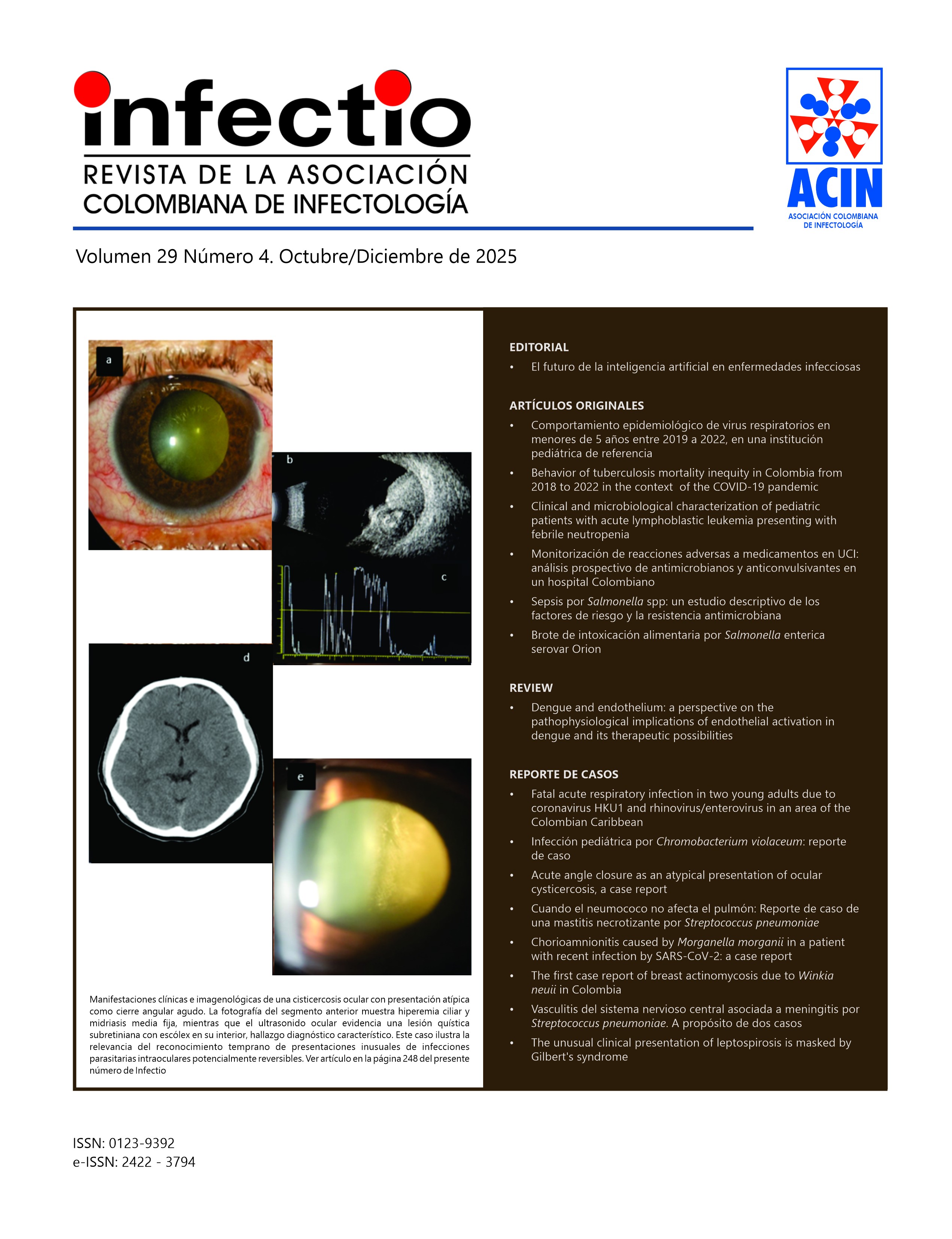
La cisticercosis ocular es una enfermedad endémica de países en vías de desarrollo, con presentaciones clínicas diversas según la localización del quiste. Se destaca un caso de cisticercosis intraocular subretiniana que debutó con un cierre angular agudo, una presentación clínica atípica. El tratamiento inicial involucró terapia médica con albendazol, esteroides orales e hipotensores oculares. En el seguimiento, a pesar del control de la sintomatología y la hipertensión ocular, se evidenció una importante reacción inflamatoria intraocular que requirió manejo quirúrgico. Se realizó una vitrectomía vía pars plana con un desenlace anatómico y visual favorable. Este caso resalta la importancia del reconocimiento de presentaciones clínicas inusuales para favorecer el diagnóstico y tratamiento oportuno, y sugiere que el tamaño del quiste puede influir en la respuesta inflamatoria y en la elección del manejo médico o quirúrgico.
Detalles del artículo
Citas
Pujari A, Bhaskaran K, Modaboyina S, Das D, Saluja G, Samdani A, et al. Cysticercosis in ophthalmology. Surv Ophthalmol. 2022 MarApr;67(2):544-9. PMID: 34339720. https://doi.org/10.1016/j.survophthal.2021.07.002
Ganesh SK, Priyanka. Analysis of Clinical Profile, Investigation, and Management of Ocular Cysticercosis Seen at a Tertiary Referral Centre. Ocul Immunol Inflamm. 2018;26(4):550-7. PMID: 29308965. https://doi.org/10.1080/09273948.2017.1413395
Sharma T, Sinha S, Shah N, Gopal L, Shanmugam MP, Bhende P, et al. Intraocular cysticercosis: clinical characteristics and visual outcome after vitreoretinal surgery. Ophthalmology. 2003 May;110(5):996-1004. PMID: 12750103. https://doi.org/10.1016/S0161-6420(03)00096-4
Rodríguez-Morales AJ, Yepes-Echeverri MC, Acevedo-Mendoza WF, Marín-Rincón HA, Culquichicón C, Parra-Valencia E, et al. Mapping the residual incidence of taeniasis and cysticercosis in Colombia, 2009-2013, using geographical information systems: Implications for public health and travel medicine. Travel Med Infect Dis. 2018 Mar-Apr: 22:51-7. PMID: 29288739; PMCID: PMC5940541. https://doi.org/10.1016/j.tmaid.2017.12.006
Otranto D, Eberhard ML. Zoonotic helminths affecting the human eye. Parasit Vectors. 2011 Mar 23; 4:41. PMID: 21429191; PMCID: PMC3071329. https://doi.org/10.1186/1756-3305-4-41
Chandra A, Singh MK, Singh VP, Rai AK, Chakraborty S, Maurya OP. A live cysticercosis in anterior chamber leading to glaucoma secondary to pupillary block. J Glaucoma. 2007 Mar;16(2):271-3. PMID: 17473746. https://doi.org/10.1097/IJG.0b013e31802d6dc2
Ratra D, Phogat C, Singh M, Choudhari N. Intravitreal cisticercosis presenting as neovascular glaucoma. Indian J Ophthalmol. 2010 JanFeb;58(1):70-3. https://doi.org/10.4103/0301-4738.58478
Lim WK, Chee SP. Nonsurgical management of subretinal cysticercosis. Retina. 2004 Jun;24(3):469-71. PMID: 15187678. https://doi.org/10.1097/00006982-200406000-00026
Singh J, Singh R. Submacular Parasite Masquerading as Posterior Pole Granuloma. Case Rep Ophthalmol Med. 2015; 2015:910383. https://doi.org/10.1155/2015/910383
García Franco R, Arias Gómez A, Guzman Cerda J, García Roa M, Ramirez Neria P. Submacular Cysticercosis Successfully Treated through Conservative Management: Case Report. Case Rep Ophthalmol. 2020 Jul 7;11(2):315-21. PMID: 32774298; PMCID: PMC7383207. https://doi.org/10.1159/000508030